
【Disclaimer】The images and text are sourced from the internet. If there is any infringement, please contact us for removal!
Note: The audio is read by a robot. If the experience is not satisfactory, those with high demands may choose not to listen. Thank you for your understanding.
The sclera, or the white part of the eye, is normally milky white and opaque. In infants, the sclera is thinner, which can cause a bluish-white appearance due to pigment transparency. In elderly individuals, the sclera may appear light yellow due to fat deposits.
The thin membrane covering the eyeball is called the bulbar conjunctiva, while the membrane covering the inner eyelid is called the palpebral conjunctiva. The junction of these two conjunctivae is known as the conjunctival fornix.
The scleral diagnosis method primarily involves observing changes in the blood vessels (luo mai) between the sclera and conjunctiva, as well as any abnormal phenomena such as black spots, blue spots, or petechiae, to infer systemic diseases.
During examination, the examiner should use their fingers to hold open the subject’s eyelids, exposing the sclera and conjunctival fornix. When examining the upper part of the sclera, the subject should look towards their toes, and when examining the lower part, they should look towards their head to fully expose the area being examined.
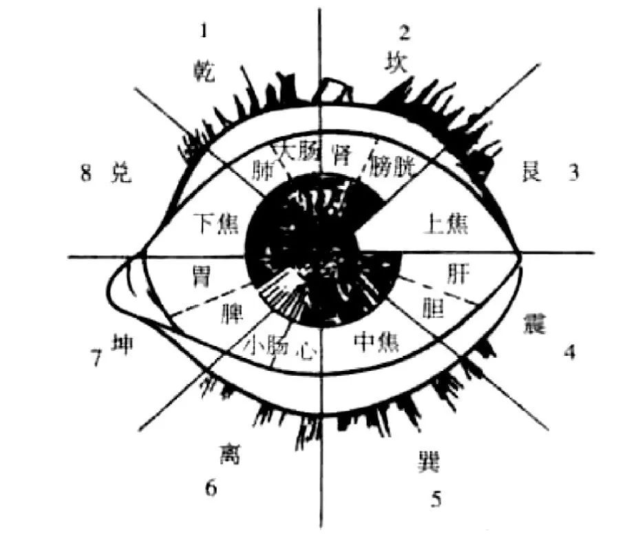
Division of the Eye Area Sclera and Its Relation to Internal Organs
The eyes are connected to the five internal organs. By dividing the eye into eight zones using the Bagua (Eight Trigrams), we can determine the corresponding reflection areas of the organs in the eyes, which has certain guiding significance for diagnosing and treating diseases.
Taking the left eye as an example, for convenience, the trigrams Qian, Kan, Gen, Zhen, Xun, Li, Kun, and Dui are represented by the numbers 1 to 8.
The specific method is as follows:
Both eyes should be looking straight ahead, drawing a horizontal line through the center of the pupil that extends past the inner and outer canthus, and then drawing a vertical line through the center of the pupil that extends past the upper and lower eyelids. This divides the eye into four quadrants, and each quadrant is further divided into two equal areas, resulting in eight equal zones, which represent the eight meridian areas.
When dividing the zones, the person should lie down with their head facing north and feet facing south. The northwest of the left eye corresponds to the Qian trigram, north corresponds to Kan, northeast to Gen, east to Zhen, southeast to Xun, south to Li, southwest to Kun, and west to Dui.
In relation to the internal organs, Qian corresponds to metal, which generates water (Kan), associated with the kidneys and bladder; water generates wood (liver and gallbladder in the east); wood generates fire (heart and small intestine in the south); fire generates earth (Kun in the southwest, spleen and stomach). Gen in the northeast represents mountains (upper jiao), Xun in the southeast represents wind (middle jiao), and Dui in the west represents marshes (lower jiao).
The eight zones of the left eye are illustrated below:
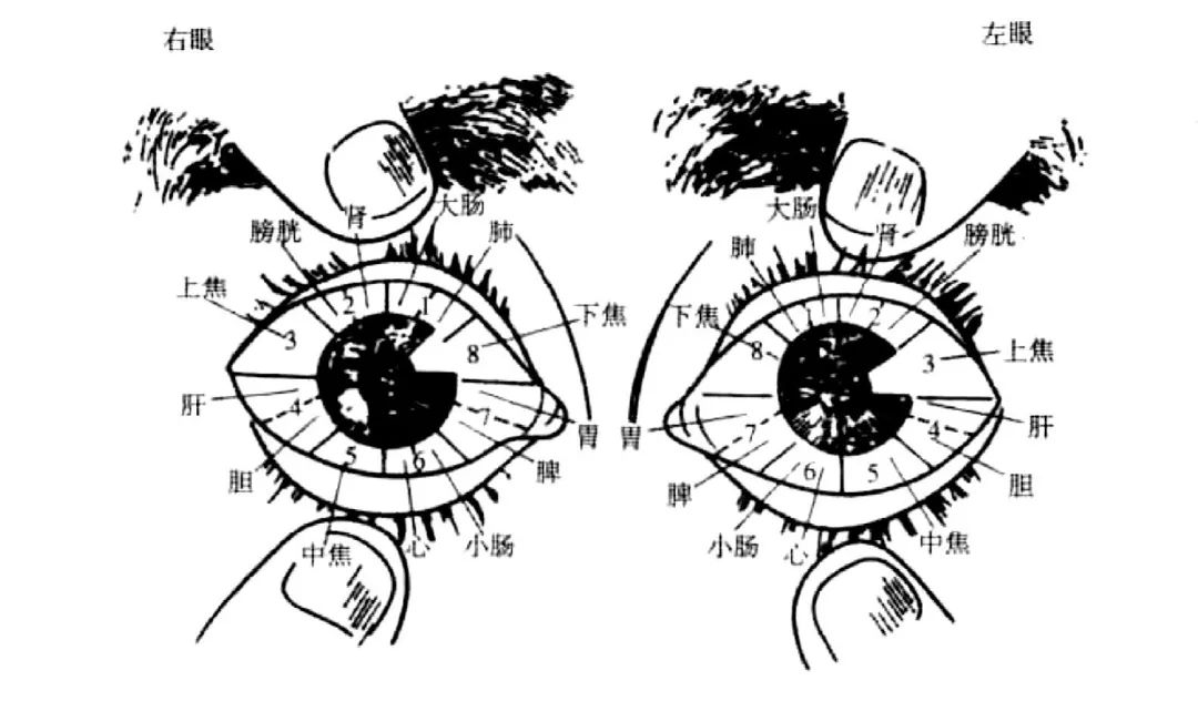
According to the theory of meridian distribution in Traditional Chinese Medicine, the meridians in the human body are symmetrically distributed, except for the Ren and Du meridians which run along the anterior-posterior midline. Thus, the outer side of the left eye corresponds to the outer side of the right eye, and the inner side corresponds to the inner side. Therefore, we can flip the left eye diagram to represent the eight zones of the right eye, as shown below:
Practical experience has shown that this method has a high accuracy rate for diagnosis and treatment.
Abnormal Spots on the Sclera
1. Blue spots appear on the sclera as irregular, non-protruding blue and purple-brown spots, ranging from the size of a pinhead to a mung bean. The edges of the spots are often clear, but can also be blurred, with a number ranging from 1 to 7. This sign is commonly seen in patients with intestinal ascariasis.
2. Black spots are found at the tips and adjacent areas of capillaries between the sclera and conjunctiva, appearing as one or more dark blue-black spots, with a diameter of 1 to 3 millimeters. This sign often indicates the presence of ascaris or pinworm infection.
Large black spots indicate the presence of adult parasites; small black spots indicate larvae. A greater number of black spots suggests a higher parasite load, while fewer black spots indicate a lower load.
Black spots typically appear in the scleral area above the left and right pupils, and may also be seen below. Therefore, during examination, the patient can be asked to move their eyes up, down, left, and right for a comprehensive observation.
3. Purple cloud spots appear as variably shaped light purple, cloud-like patches at the tips and edges of capillaries between the sclera and conjunctiva.
This sign suggests the patient has hookworm infection, with larger patches indicating a more severe infection, and smaller patches indicating a milder infection.
It is important to differentiate purple cloud spots from blue spots, and this can be assessed in conjunction with the patient’s medical history and symptoms.
4. Petechiae, also known as “report injury spots,” appear as raised blue-purple small blood vessels on the sclera or between the sclera and conjunctiva, with the ends of the vessels appearing darker, resembling pinhead-sized dots.
These petechiae, in the absence of any eye disease or subjective symptoms, often indicate that some part of the body has sustained an injury.
If the petechiae are located away from the ends of the blood vessels and are nearby or in the middle, they have no diagnostic value.
The color and shape of the petechiae can help determine the extent of the injury.
For example, if the color is light like clouds or black with white, scattered and not clustered, it indicates an injury in the Qi level; if the color is black and dense, resembling small sesame seeds, it indicates an injury in the blood level; if the area around the black spots is light like clouds and irregularly shaped, it indicates injury to both Qi and blood.
Based on the location of the petechiae on the eye, one can determine the site of injury in the body. Through extensive clinical observation, the following patterns have been identified:
(1) If the report injury spots appear in the upper half of the eye, they primarily reflect injuries to the waist, back, and upper limbs; petechiae in the waist area tend to be more medial or closer to the pupil; those in the scapula and spine are often centered; small blood vessels in the upper limbs are short, with petechiae tending to be more lateral and farther from the pupil; in the lower limbs, small blood vessels are longer, thus exceeding the horizontal line of the pupil; if both upper and lower limbs are injured, the small blood vessels may appear interrupted.
(2) If the report injury spots appear in the lower half of the eye, they primarily reflect injuries to the chest and lower limbs. Injuries above the nipple will have petechiae centered; injuries on the inner side of the nipple or beside the sternum will have petechiae more medial; injuries on the outer side of the nipple, below, and under the clavicle will have petechiae more lateral; injuries on both sides of the sternum will show a “Y” shaped bifurcation of small blood vessels, with petechiae located at the ends of the bifurcation.
(3) If the report injury spots appear on the outer side of the eye, the order of petechiae from top to bottom corresponds to injuries along the posterior axillary line, mid-axillary line, and anterior axillary line. If they appear on the inner side of the eye, it suggests injury to the opposite side of the axilla.
(4) Some small blood vessels connected to the petechiae may appear engorged in a spiral shape, while others may appear wavy, hooked, or of varying thickness.
The spiral shape suggests severe pain and indicates symptoms of both Qi and blood injury; the wavy shape suggests neuralgia; if the blood vessels suddenly bend at an angle, it indicates reflex pain; hooked blood vessels indicate a shift to the opposite side; and blood vessels of varying thickness, even without petechiae, also suggest injury.
(5) In addition to diagnosing injuries, some petechiae can also indicate other diseases.
For example, if the ends of the blood vessels reach the bulbar conjunctiva, and the petechiae are black-red or pink and relatively large, it suggests liver disease.
If there are bright red or black, blue spots on the outer side of both eyes, it suggests kidney disease.
Additionally, if green spots appear on the sclera, it often indicates intestinal obstruction.
Changes in Capillaries on the Sclera
On the sclera, one can see faintly intersecting capillaries, known in Traditional Chinese Medicine as luo mai.
In normal individuals, the capillaries are fine and not prominent, especially in children’s eyes. If they have not suffered from major illnesses, the sclera is often clean and bluish-white, with no visible capillary distribution.
However, if illness occurs, or if the skin transmits through the meridians to the internal organs, or if the internal organs transmit to the skin, regardless of whether one or several meridians are affected, it can be revealed on the sclera. Once capillaries appear, their traces and growth are not easily erased.
There are generally seven types of capillary conditions on the sclera:
1. Thick roots.Capillaries at the edge of the white part of the eye are thick and gradually thin out towards the front; this shape often indicates chronic diseases.
2. Varicosity or engorgement.Capillaries appear engorged, extending from the roots, with bends and twists, indicating a more severe condition.
3. Extension.Capillaries extend from one meridian area to another, indicating that the disease has shifted from one organ to another; or the disease in the latter organ is caused by the former organ, indicating the source of the disease.
4. Multiple bifurcations.This phenomenon often occurs in the upper part of the eyeball, and sometimes in the lower part, indicating that the disease is unstable and prone to change.
5. A raised line.This often indicates diseases of the six fu organs, as the capillaries of the sclera and conjunctiva differ in depth; diseases of the five zang organs often appear deeper, resembling capillaries beneath a glass plate, while diseases of the six fu organs often appear on the surface, resembling capillaries on top of a glass plate.
6. A small blurred area.This type of capillary often occurs in the liver and gallbladder area, commonly seen in liver qi stagnation and gallstones.
7. Dripping.The lower ends of the capillaries on the sclera appear to be dripping like dew drops. This has been previously described and is often seen in gastrointestinal issues, commonly indicating parasitic accumulation; in other meridian areas, it often indicates stagnation.
The colors of the capillaries on the sclera can generally be categorized into eight types:
1. Bright red capillaries indicate a new disease, belonging to excess heat, and the disease is currently developing.
2. Purple-red indicates a condition of excessive heat.
3. Dark red indicates a heat disease with worsening condition.
4. Red with black indicates heat disease penetrating inward.
5. Red with yellow indicates the disease is alleviating.
6. Light yellow indicates the disease is about to heal.
7. Pale indicates deficiency of Qi and blood, belonging to deficiency or cold syndrome.
8. Dark gray indicates chronic lesions, suggesting that the disease has healed; if it changes from dark gray to light red, it is a sign of recurrence of an old disease.
If there are phenomena such as congestion, varicosity, thickening, color changes, and abnormal extensions of capillaries at different locations between the sclera and conjunctiva, it often indicates that some internal organ in the body has undergone pathological changes.
Common conditions include the following nine situations:
1. The small blood vessels in the lower conjunctiva appear wavy, often indicating heart disease.
2. The small blood vessels on the inner side of the upper conjunctiva reaching the bulbar conjunctiva appear purple-red and relatively thick like straight lines, indicating spleen disease.
3. On the inner side of the upper conjunctiva, if there are vesicles resembling herpes on or around the blood vessels, it often indicates lung disease.
4. Between the sclera and conjunctiva, directly below the pupil (at the 6 o’clock position), if small blood vessels are congested and dilated, it often indicates stomach disease or hyperacidity.
5. In the inner lower area of the eye, if small blood vessels between the sclera and conjunctiva are congested and dilated, appearing light blue, it often indicates hepatitis.
6. If there is a blood vessel on the sclera that appears in a straight line, crossing from the inner to the outer corner of the eye, if it occurs in the left eye, it often indicates stomach disease; if it occurs in the right eye, it often indicates liver disease; this phenomenon can also be seen in individuals who frequently consume white liquor.
7. If congested blood vessels enter the pupil from the sclera, it is referred to as “red vein penetrating the pupil,” which is a sign of scrofula (cervical lymph node tuberculosis).
If only one red vein penetrates the pupil, it indicates a mild condition; if 2-3 red veins penetrate simultaneously, it indicates a severe condition; if the red veins do not penetrate the pupil, it indicates the mildest condition.
8. If the upper half of the eyeball shows horizontal blood vessels (healthy individuals show a “人” shaped pattern), causing the blood vessels to appear U-shaped, or if the sub-conjunctival layer shows a straight line, it can be seen in malignant tumors of the digestive tract, such as gastric cancer, intestinal cancer, liver cancer, and esophageal cancer.
9. In the outer lower area of the eye, between the sclera and bulbar conjunctiva (between the 5 o’clock and 6 o’clock positions), if small blood vessels are thick, significantly congested, and dilated, with colors ranging from bright red, light red, or red with yellow or black, it often indicates hemorrhoids. This phenomenon is referred to as scleral hemorrhoid sign.
If one hemorrhoid sign appears without branching at the end, it indicates only one hemorrhoid; if there are branches at the end, or if two hemorrhoid signs appear at the same location, it indicates two hemorrhoids; a greater number of hemorrhoid signs or more branches indicate a higher number of hemorrhoids.
If the hemorrhoid signs are small, not very engorged, and not prominent, it indicates small hemorrhoids; if the hemorrhoid signs are thick and engorged, it indicates large hemorrhoids; if the root of the hemorrhoid sign is particularly swollen, or if several are clustered together, it indicates prolapsed hemorrhoids.
Hemorrhoid signs appearing in the left eye indicate internal hemorrhoids on the left side of the anus; conversely, those in the right eye indicate the right side; if both eyes show signs, it indicates hemorrhoids on both sides.
The aforementioned methods may lose diagnostic significance if there are other eye diseases or conditions such as trachoma, acute or chronic conjunctivitis, photokeratitis, increased intracranial pressure, and pulmonary diseases, which can cause congestion and dilation of the conjunctival blood vessels.
Additionally, this hemorrhoid sign is limited to internal hemorrhoids; it has no significance for external hemorrhoids or anal fissures.
If there are numerous and disordered blood vessels in the aforementioned areas, based on the theory of the lung and large intestine being interrelated, one should consider the possibility of lung or bronchial lesions, which cannot be interpreted as hemorrhoid signs. The hemorrhoid signs should also be compared with signs of gastric disease and hepatitis.
Note:Specific treatments and medications should be followed under medical guidance!This article is excerpted from “Strange Methods of Diagnosis,” compiled by Zhao Jiancheng.
Source:“Left Hand for Cold Damage, Right Hand for Warm Disease”
Highlights from Previous Articles
There is a form of emotional violence called “I am only doing this for your good.”
Judgment is the root of all suffering.
How to maintain a sense of security in intimate relationships?
Ten experiences: Those without a strong inner self are not qualified to talk about life.
Discovering the deep wounds within can lead to true maturity.
Why can cultivating the mind heal diseases?
The “Stretching and Bone-Pulling Walking Method” that can help people grow taller.
Women’s health: Important lifestyle habits, can you wash your hair during menstruation?
The most famous pediatrician in TCM history: Qian Yi (Part 2) | Luo Dalun
Academician of the Chinese Academy of Sciences reveals the truth about hypertension and antihypertensive drugs.
Hypertension, stroke, cerebral hemorrhage, cerebral thrombosis | Qu Limin
“Common Knowledge for Treating Diseases” 37 points.
Secret recipes that even doctors praise.
The most important moment is always now.
Condensing the wisdom of practice into six key principles.
Reading texts aloud can change a child’s character; the benefits are numerous.
Good people have a spiritual light above them.
A story about dignity – The oil tycoon Hammer.
Complete Guide to the Diagnosis of Blue Veins
Inquiry on Classical Formulas: How to Conduct Effective and Practical Inquiry on Classical Formulas?
Information on 60 well-reputed TCM practitioners in Chengdu, Sichuan, including “Six Little Children,” Feng Daka, Huang Yizhen, Chen Fuzhi, Lu Huoshen, etc. Each possesses unique skills (Good TCM practitioners).
100 diagnostic experiences, medication experiences, and formula experiences from veteran TCM practitioners.
60 secrets of effective acupuncture points for diagnosis and treatment: By feeling the tender points, you can know what disease it is; by finding the acupuncture points, you can solve the pain yourself.
[Spinal Diagnosis Collection] 108 types of internal organ diseases originating from “Spinal Misalignment” with corresponding illustrations.
One must be a TCM practitioner skilled in inquiry.
TCM pulse diagnosis is so magical; it is truly incredible.
Self-learning pulse diagnosis (pulse examination) should start with distinguishing strength and weakness, and then from speed to cold and heat.
Wang Xingfu’s medical discussions – Discussing the application of tongue and pulse signs in diagnosis.
The significance of pulse diagnosis – Qualitative, locational, quantitative, and situational.
Without pulse examination and inquiry, how does Mr. Dong Jingchang diagnose? (Dong’s Special Acupuncture Points).
Divided Meridian Pulse Diagnosis – Restoring the Comprehensive Examination Method of the “Huangdi Neijing”.
Practical Experience in Cold Damage: How to Effectively Improve Diagnostic Accuracy?
Xu Yueyuan: The unity of pulse and person.
Why can pulse diagnosis reveal diseases?
What does sweat tell you? Inquiry in TCM regarding “sweat”.
What does pain tell you? Inquiry in TCM regarding “pain”.
This article is: The Shortcut to TCM Four Diagnoses.
Inquiry Thinking: A Detailed Explanation of TCM Thinking Around Inquiry.
Insights from Following Master Liu Duzhou.
Master of TCM Zhou Zhongying: Insights and Techniques from 60 Years of TCM Diagnosis.
Complete Guide to Blue Vein Diagnosis: Illustrated Diagnosis of Common Blue Veins in the Human Body.
Precious TCM Examination: Clinical Revelation of Shadi Diagnosis.
“The Clear Insights of Pulse Diagnosis” – A Collection of Insights from Pulse Diagnosis.
Help you summarize: A complete list of abdominal diagnosis in cold damage (illustrated version).
Log of Pulse Diagnosis by Folk Experts, with significant practical reference value.
Folk Experts: Observing Tongue Diagnosis from the Perspective of Qi Mechanism (True Tongue Diagnosis).
Insights Gained from Following a Master for a Day – 45 – Dream Diagnosis.
Detailed Explanation of TCM Inquiry Methods by Ni Haixia.
The Soul and Mystery of TCM Diagnosis | Meng Jingchun.
Ear Diagnosis.
Key Points of Hand Diagnosis Illustrated in “Unique Hand Diagnosis”.
Special Features of Ear Diagnosis by Zhu.
The Most Comprehensive: Diagnostic Songs (Collection).
Inquiry! How many truly master inquiry?
Essence of Zang-Fu Diagnosis: A Quick Start Guide.
Complete Collection of Abdominal Diagnosis (Three Great Experts, Three Great Sections).
Niuwen: Diagnosing and Treating Diseases with Ten Fingers.
Simply Self-Learning TCM Series One: Disease Mechanism Diagnosis.
Strange Talks on Diagnosis: Foot Diagnosis Method.
Strange Talks on Diagnosis: Leg Diagnosis Method.
“Children are mute, observation is key” (Essence of Pediatric Observation).
This is the true “Ancient TCM Tongue Diagnosis” (Preserved for careful reading).
Four Diagnostic Methods in Gynecology, Complete Explanation of Gynecological Differentiation.
Excellent Foundation of TCM Observation.
These 22 types of hand diagnosis illustrations are considered “Perfect Diagnostic Diagrams”.
Special Diagnostic Methods: Detailed Explanation of Sweat Diagnosis.
Complete Guide to Cold and Heat Inquiry in TCM | Zhu Wenfeng.
TCM Teaches You Pulse Diagnosis (Collection).
TCM Experts Share Their Learning Experiences in Pulse Diagnosis…
Pulse Diagnosis Essentials: The Four-Word True Formula of TCM Pulse Diagnosis (Including: Twelve Simple Pulse Diagnosis Methods).
Observation and Understanding of TCM Diagnosis – The Four Diagnostic Methods of TCM.
TCM Diagnosis Secrets – The Mystery of Observation, Inquiry, Auscultation, and Palpation.
Senior TCM Practitioners’ Complete Inquiry Guide, Revealed!
Is TCM really that simple? (Including Inquiry Form).
TCM practitioners skilled in inquiry are superhumans with multiple roles!
Ni Haixia: Inquiry in Classical Formulas is Very Important | Detailed Explanation of Inquiry Methods.
Ni Haixia: Diseases are inquired about | Detailed Explanation of Inquiry Methods.
How do veteran TCM practitioners take pulses?
The Thought Process of TCM Pulse Diagnosis – Teaching You How to Take Pulses.
What is the experience of pulse diagnosis? Teacher Dan Fuzai shares his pulse-taking insights.
My Pulse Diagnosis Experience: Ten Symptoms that Reveal Cold Damage Pulse Patterns (Recommended Reading).
Common Symptoms and Pulse Diagnosis of Classical Formulas.
[Zou Mengcheng] Learning to Observe Pulses and Experiences in Pulse Diagnosis.
Pulse Diagnosis Basics (Including: Differentiation of Similar Pulses, Seven Unique Pulses).
Pulse Diagnosis for Pregnancy: A Skill You Can Master.
Pulse Diagnosis Techniques.
TCM’s Unique Skills: The Secret Pulse Diagnosis that Can Predict Diseases!
Senior TCM Practitioners Discuss Pulse Diagnosis – Teaching You to Quickly Master Pulse Diagnosis!
Pulse Diagnosis Basics: Learn to Take Pulses in Ten Minutes!
Introduction to TCM Pulse Diagnosis | Learn to Understand Pulse Diagnosis in Ten Minutes!
He lost his sight at 9, and after 40 years of feeling countless pulses, he uses mathematics and fluid mechanics to explain the science of pulse diagnosis.
========== END ==========
Warm Reminder: This platform shares health-related images and information for reference and learning purposes only and should not be used as a basis for medical diagnosis. If needed, please use under the guidance of a physician.
⊙ Copyright Statement: The article is sourced from the internet. If there is any infringement, please contact us for removal.
Advertisements are randomly assigned by the system, and we cannot control or select them in detail. Please do not purchase any animal products or meat. Do not buy fishing or hunting equipment, refuse to kill; do not play violent games, as they are fundamentally the same as actual killing; do not read pornographic articles, as all evil stems from lust, and sexual misconduct is the root of reincarnation and disaster. Cause and effect follow closely. Remember!
As parents, not knowing medicine is unkind! As children, not knowing medicine is unfilial!
Can’t find a good TCM practitioner? Why not learn TCM yourself!
In the menu of this account, there is a complete series for beginners and a video series. Everyone is welcome to learn.
Learning Ancient TCM, Inheriting the Classical Formulas of Cold Damage.
Virtue carries all things! Grass, trees, metals, and stones can only remove physical ailments; cultivating oneself and nurturing virtue is the way to eliminate inner demons!
Wishing: Peace and harmony in the world, clear sun and moon; timely wind and rain, no disasters; a prosperous nation and peaceful people, no need for weapons; promoting virtue and kindness, diligently practicing propriety; no thieves in the country, no grievances; the strong do not bully the weak, everyone gets their due; no one suffers from illness, wealth and health; longevity and good virtue, starting and ending well.