Observation of Collaterals
1. Observation of Pediatric Index Finger Collaterals
The method of observing the index finger collaterals in children first appeared in the Tang Dynasty in Wang Chao’s “Shuijing Tujue”. This method developed from the collateral diagnosis of the fish border in the “Lingshu” and holds significant diagnostic value for children under three years old. The collaterals on the inner side of the index finger are also branches of the Taiyin Hand Meridian (Taiyin Shou Mai, 手太阴之脉), which runs from the chest to the hand, reaching the fish border and extending to the tip of the thumb. The branches emerge from the wrist and run along the inner edge of the second finger, thus the diagnosis of the index finger collaterals is analogous to that of the fish border collaterals and the cun, guan, chi pulses. Due to the small size of children’s pulses and their tendency to cry and move during diagnosis, the accuracy of pulse diagnosis can be affected. However, because children’s skin is thin and delicate, the collaterals are more prominent, making the observation of collaterals more convenient than pulse diagnosis.
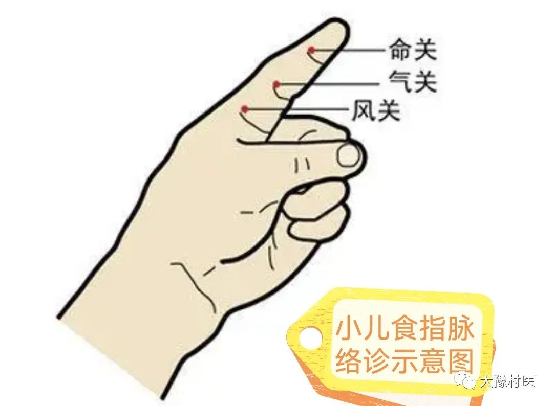
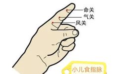
(1) Location: The manifestation and distribution of the index finger collaterals can be divided into three sections: wind, qi, and life. The first section of the index finger is the wind section, which is between the distal transverse crease of the metacarpophalangeal joint and the first transverse crease; the second section is the qi section, which is between the second and third transverse creases; the third is the life section, which is from the third transverse crease to the tip.
(2) Method of Diagnosing Collaterals: Hold the child towards the light, the physician uses the left hand to grasp the child’s index finger, and with the right thumb applies moderate pressure from the life section towards the qi and wind sections, pushing several times. The collaterals become more pronounced, facilitating observation.
(3) Differentiating Severity in the Three Sections: When external pathogens invade the body, they often penetrate from superficial to deep, first entering the collaterals, then the meridians, and finally the organs. The appearance and color of the collaterals change according to the depth of the invasion. If the collaterals are prominent in the wind section, it indicates that the pathogen has entered the collaterals, suggesting a mild condition. If the collaterals are visible from the wind section to the qi section, their color is darker, indicating that the pathogen has entered the meridians, suggesting a more severe condition. If the collaterals are prominent in the life section, it indicates that the pathogen has penetrated the organs, which may be life-threatening, hence it is called the life section. If the collaterals reach the tip of the finger, it is termed “through the section and piercing the armor,” indicating a more dangerous condition with a poor prognosis. The diagnostic method for internal injuries and miscellaneous diseases also follows the principle that collaterals visible in the wind section indicate a mild condition, those in the qi section indicate a more severe condition, and those in the life section indicate the most severe condition.
(4) Shape and Color Indicating Disease:
① Normal Shape and Color: The normal color of the collaterals is light red, a mix of red and yellow, subtly visible within the wind section; they are mostly not prominent, and may even be inconspicuous, often appearing slanted, single-branched, and of moderate thickness. However, the thickness can also be related to climatic conditions; heat causes them to thicken and lengthen, while cold causes them to thin and shorten. The length also correlates with age, being longer in children under one year and shortening with age.
② Floating and Sinking: Floating collaterals indicate that the disease is superficial, often seen in exterior pathogenic conditions. Sinking collaterals indicate that the disease is internal, commonly seen in both exterior and internal conditions. However, clinical observations show that healthy children can also have slightly floating or sinking collaterals.
③ Depth: Darker colors indicate more severe disease, while lighter colors indicate milder disease; pale colors suggest deficiency, while stagnant colors suggest excess. In cases of yin and yang collapse, the yang does not reach the extremities, resulting in a pale color that may not be visible. If the pathogen invades the heart and causes closure, it often leads to stagnation of qi and blood, resulting in dark and stagnant collaterals.
④ Color: Purple-red color indicates internal heat; bright red indicates exterior pathogenic conditions; blue indicates wind and various pain conditions; pale color indicates deficiency; purple-black color indicates blood stasis, a sign of critical illness.
⑤ Shape: Collaterals that gradually lengthen indicate disease progression and worsening; those that shorten indicate disease regression and improvement. However, there are cases of fluid depletion and dual deficiency of qi and yin, where the collaterals shorten below the wind section due to insufficient qi and blood. Yin deficiency with floating yang often presents with elongated collaterals.
Collaterals that become thicker are often associated with heat or excess conditions; those that become thinner are often associated with cold or deficiency conditions. Single-branched and slanted collaterals are often associated with mild disease; curved, circular, or multi-branched collaterals indicate severe disease, often associated with excess conditions.
2. Observation of Fish Border Collaterals
The fish border is the fleshy area behind the base of the thumb. The fish border belongs to the Taiyin Lung Meridian (Taiyin Fei Jing, 手太阴肺经), and the principle of diagnosing the fish border collaterals is consistent with the principle of pulse diagnosis at the cun position. Additionally, the qi and blood in the collaterals are derived from the spleen and stomach, with stomach qi rising to the Taiyin, hence the diagnosis of fish collaterals can also reflect stomach qi.
The “Lingshu: Meridian Chapter” states: “In diagnosing collaterals, if the pulse color is blue, it indicates cold and pain; if red, it indicates heat.” Cold causes qi and blood to congeal, leading to a blue-black color; heat causes qi and blood to become turbid, leading to a yellow-red color. Therefore, if there is cold in the stomach, the cold qi reaches the fish border, resulting in blue collaterals; if the collaterals are blue and small, it indicates qi deficiency, which is a deficiency condition. If there is heat in the stomach, the heat qi reaches the fish border, resulting in red collaterals. The “Four Diagnostic Methods” summarizes: “More red indicates more heat; more blue indicates more pain; more black indicates long-standing numbness; red, black, and blue colors often indicate cold and heat.”
3. Observation of Nail Shape and Color
Nails are the remnants of tendons and are external indicators of the liver and gallbladder. The liver stores blood and governs the smooth flow of qi, thus observing the nails can reveal the state of qi and blood circulation.
Normal nails are rosy, firm, and arc-shaped with a sheen; when pressed at the tip, the color quickly returns upon release, indicating sufficient qi and blood and smooth circulation.
If the nail bed is deep red, it indicates heat in the qi level; yellow indicates jaundice, often due to damp-heat; pale white indicates blood deficiency or dual deficiency of qi and blood; pale color indicates deficiency-cold, often due to spleen and kidney yang deficiency; purple-black indicates blood stasis or blood coagulation; blue indicates cold conditions.
If pressing the nail turns white and the color returns slowly upon release, it indicates blood stasis or qi stagnation; if it does not return to red, it often indicates blood deficiency. Flat and concave nails, known as “reversed nails,” often indicate liver blood deficiency. Dry and brittle nails indicate bi syndrome or bone pain. Pale and brittle nails indicate liver heat.

Email: [email protected]