Here are commonly seen clinical tongue diagnosis charts, presented for those who love Traditional Chinese Medicine (TCM).

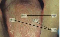
Tongue Diagnosis Organs Location Chart
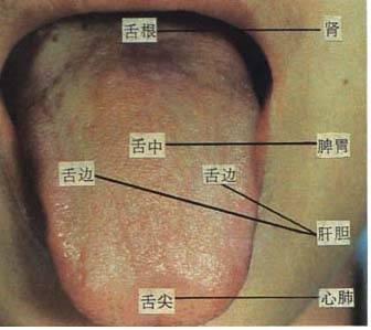
According to the “Neijing” (Inner Canon), different areas of the tongue can reflect pathological changes in various organs, which has certain reference significance in clinical practice. The tip of the tongue corresponds to the upper jiao (heart and lungs); the middle part corresponds to the middle jiao (spleen and stomach); the root corresponds to the lower jiao (kidneys); the sides correspond to the liver and gallbladder.
The “Shanghan Zhi Zhang · Cha She Bian Zheng Fa” also states that “the tip of the tongue belongs to the upper jiao, the middle part belongs to the middle jiao, and the root belongs to the lower jiao”.
Tongue Color Section
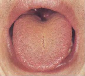
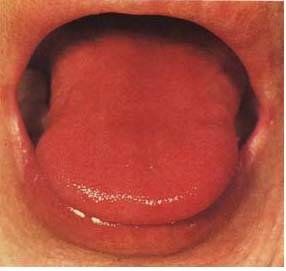
1. Normal Tongue
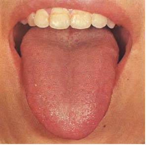
The characteristics of a normal tongue are: a light red color, moist texture, moderate size, and flexibility; the tongue coating is even, thin, white, and moist, referred to as “light red tongue, thin white coating“.
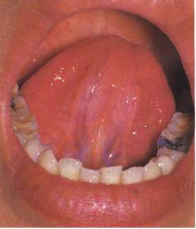
The normal sublingual vessels are only faintly visible, appearing as purple-red lines, and should not be swollen. Most are single branches, with the largest diameter not exceeding 2.7mm, and the length generally does not exceed 3/5 of the line connecting the tip of the tongue to the sublingual tubercle.
2. Pale White Tongue

Pale white tongue is often seen in cases of yang deficiency with excess cold, and qi deficiency with blood deficiency. This tongue appears pale white, with a thin white and moist coating, indicating deficiency of both qi and blood.
3. Withered White Tongue
The tongue color and the gums and lips show no blood color, referred to as “withered white”. This tongue appears withered and dry, lacking vitality, indicating decline of essence and qi, with a critical condition.
4. Red Tongue
Red tongue is seen in cases of external heat or yin deficiency with excess fire. This tongue appears red, with a thin yellow coating, and a rough texture, indicating excess heat in the qi level.
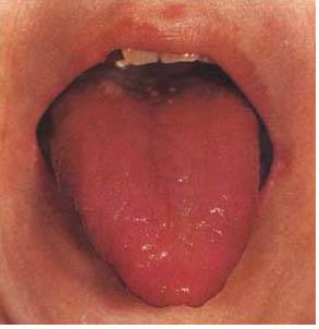
5. Red Tip of the Tongue
The red tip of the tongue is often seen in cases of heart fire rising, indicating heat in the upper jiao. This tongue has a red tip with prickles, while the rest is light red and moist, indicating excess heat in the upper jiao.
6. Deep Red Tongue (1)
The tongue is deep red, referred to as “deep red tongue”, indicating excess heat. In cases of external diseases, it signifies evil heat entering the nutrient level; in internal injuries with yin deficiency and excess fire, it is also commonly seen. This tongue appears deep red and dark, with leaf vein-like shallow cracks, and little coating, indicating excess heat, qi and blood stagnation, with damage to the righteous qi.
7. Deep Red Tongue (2)
This image is from a patient with chronic gastric fire, showing a deep red tongue, dry with no coating, and shallow cracks on the surface, indicating internal heat blazing, gastric yin depleted, and significant damage to gastric qi.
8. Deep Red Tongue (3)
The tongue is deep red, with little coating, and many red spots on the front part (i.e., fungal-like congestion), indicating excess heat, with damage to both qi and yin.
9. Cyanotic Purple Tongue
The cyanotic purple tongue is caused by stagnation of qi and blood. This image shows a patient with chronic bronchitis complicated by infection, pulmonary emphysema, and heart failure, with a dark purple tongue and white greasy coating, indicating phlegm and fluid retention, lung and kidney qi deficiency, and blood stasis.
10. Pale Purple Tongue
The pale purple tongue is often seen in cases of yang deficiency with excess yin. This image shows a patient with pulmonary heart disease, with a pale purple tongue and thin white moist coating, indicating insufficient yang qi, with poor blood circulation.
11. Pale Purple Stagnation Tongue
This image shows a patient with menstrual irregularities (dysmenorrhea), with a pale purple tongue and many purple spots, and a thin white moist coating, indicating insufficient yang qi, with cold stagnation and blood stasis.
12. Purple Red Tongue
This image shows a patient with a constitution of yin deficiency, with a red tongue and little coating, due to lung infection and heart failure, with a purple-red tongue, accompanied by fever and cough, indicating internal heat, with qi and blood stagnation.
13. Purple Red Tongue
The tongue is purple-red with prickles, indicating phlegm heat stagnation.
14. Stasis Spot Tongue
The tongue has stasis spots or stasis points, indicating blood stasis. This image shows irregularly shaped stasis spots on the sides and edges of the tongue, which is a sign of liver channel blood stasis.
Tongue Coating Section
The normal tongue coating is generated by the stomach qi and stomach yin rising to the tongue surface, while pathological tongue coating is formed by the stomach qi carrying evil qi. This chart introduces various common tongue coating changes based on coating color and texture changes.
1. Thin White Moist Coating
This image shows a normal tongue coating. The coating is moist, and through the coating, a light red tongue texture can be seen, referred to as “thin white moist coating”, indicating strong stomach qi and abundant fluids.
2. Thin White Slippery Coating
The tongue coating is excessively moist, feeling slippery upon touch, and may even drip saliva when the tongue is extended, referred to as “slippery coating”, often due to water dampness or phlegm retention. This image shows an overly moist tongue surface, with a thin white coating, slightly thicker at the root, and a plump tongue body with teeth marks on the edges, indicating insufficient yang, with water dampness retention.
3. White Greasy Coating
The greasy coating is characterized by fine and dense particles tightly adhering to the tongue surface, which do not come off when scraped, often caused by damp turbidity, phlegm retention, or food stagnation.
4. White Slippery Greasy Coating
This image shows a patient with chronic bronchitis, with a white greasy coating, excessive moisture on the tongue, pale purple tongue color, and purple spots at the tip, indicating insufficient yang, with phlegm retention.
5. White Thick Greasy Coating
The greasy coating is covered by a layer of dirty, slippery phlegm, referred to as “sticky greasy coating”, indicating excess damp turbidity, with spleen and stomach yang qi being obstructed.
6. White Thick Greasy Coating
This image shows a patient with liver cirrhosis and ascites, with a pale purple tongue and thick white greasy coating in the middle, indicating internal yang deficiency with excess yin, water dampness accumulation, and blood circulation stagnation.
7. White Thick Loose Coating
The tongue coating gradually changes from greasy to loose, with coarser particles, floating on the tongue surface, which can be scraped off, indicating yang qi recovery, gradually transforming cold dampness and yin evil.
8. White Greasy Coating Transforming to Dry
This image shows a patient with chronic bronchitis, originally with a white thick greasy coating, now most of the greasy coating has transformed to dryness, with dry cracks in the coating, and a patch of coating has fallen off, while the edges still have white coating that has not yet dried. This indicates phlegm and fluid stagnation transforming into heat, with stomach qi and fluids gradually damaged.
9. Yellow Greasy Coating Transforming to Dry
This image shows a tongue with yellow greasy coating covering the surface, with the middle and front parts of the coating gradually transforming to dryness, indicating internal heat blazing, with damp evil transforming to dryness.
10. Thin Yellow Coating
This image shows a patient with respiratory tract infection and fever. The tongue texture is slightly red, with a thin yellow coating, indicating exterior evil entering the interior, with excess heat in the qi level.
11. Yellow Greasy Coating (1)
Yellow greasy coating is often seen in cases of damp heat, phlegm heat, food stagnation transforming into heat, summer heat, damp heat, and obstructed qi in the bowels. This image shows a patient after influenza, with a light yellow thick greasy coating, chest tightness, and poor appetite, indicating damp heat obstructing the middle, with spleen losing its healthy function.
12. Yellow Greasy Coating (2)
This image shows a patient with biliary tract infection, with yellow greasy coating, indicating liver and gallbladder damp heat accumulation.
13. Yellow Greasy Coating (3)
This image shows a patient with liver cancer, with a red tongue and yellow thick greasy coating, with red spots on the tip, indicating damp evil and obstructed heat.
14. Yellow Sticky Greasy Coating
This image shows a patient with intestinal obstruction, with yellow greasy coating, and a layer of yellow sticky phlegm covering the tongue surface, caused by obstructed bowel qi, with damp turbidity rising to the tongue.
15. Yellow Slippery Coating
Yellow coating can also be seen in cold damp conditions, characterized by yellow and moist coating, with a plump tongue body, and often pale white tongue color, or with teeth marks on the edges. This indicates yang deficiency, with phlegm or cold dampness retention.
16. Yellow Dry Coating (1)
This image shows a patient with lung infection, with the tongue coating changing from white to yellow (with some white greasy coating remaining), indicating disease transforming from cold dampness to heat, with fluids already damaged.
17. Yellow Dry Coating (2)
The tongue surface is very dry, with yellow thick dry coating, rough particles, and cracks on the front part of the tongue, indicating excess heat blazing, with fluids severely damaged.
18. Yellow Petal Coating
The tongue coating is cracked into petals, yellow in color, with a red tongue and shallow cracks, indicating a sign of excess heat damaging fluids. This image shows that there is still white greasy coating on the tongue, indicating that the original pathogenic evil was phlegm, damp turbidity, and other yin evils.
19. Gray White Greasy Coating
The tongue coating changes from white greasy to gray, often seen in cases of yang deficiency with cold dampness, phlegm retention, and other yin evils accumulating in the middle jiao, which have not transformed over time.
20. Gray Yellow Greasy Coating
This image shows a patient with pulmonary heart disease, with gray yellow greasy coating and dark purple tongue color, indicating damp turbidity and phlegm heat transforming into heat, with poor blood circulation.
21. Dirty Gray Greasy Coating
The tongue coating is greasy and sticky, gray yellow in color, with a layer of dirty substance on the tongue surface, referred to as “dirty greasy coating”, indicating excess damp turbidity.
22. Black Greasy Coating
This image shows a patient with lobar pneumonia, with the tongue coating changing from white greasy to black greasy, with a red tongue, indicating internal heat blazing, with damp turbidity transforming into fire.
23. Charred Black Coating
This image shows a patient with biliary tract infection, with a red and dry tongue, with black dry coating at the root, and a little white greasy coating on the sides, indicating excess heat, with damp turbidity transforming into dryness damaging fluids.
24. Black Dry Coating
This image was taken two days before the death of a patient with gastric cancer, with a black dry coating, with black coating at the root resembling black hair, and a shriveled tongue body, indicating depletion of yin fluids, with extreme internal heat.
25. Coating Loss
Where the coating has fallen off, the tongue surface is smooth and without coating, referred to as “coating loss”, indicating deficiency of stomach yin and stomach qi, or insufficient qi and blood, unable to nourish and regenerate new coating.
26. Greasy Coating with Partial Loss (1)
This image shows a patient with pulmonary heart disease and heart failure, originally with a white greasy coating, now with partial loss on the tongue middle and front, while the sides still have yellow white greasy coating, with a cyanotic purple tongue, indicating phlegm heat obstructing, with poor qi and blood circulation, and weakened stomach qi.
27. Greasy Coating with Partial Loss (2)
The tongue coating is thick and greasy, with coating loss, indicating phlegm and dampness have not transformed, with stomach qi already damaged, indicating a mixed condition of deficiency and excess.
28. Similar Coating Loss
This image shows a patient with gastric ulcer, with multiple areas of coating loss, where the lost areas still have new papillae, referred to as “similar coating loss”, indicating insufficient qi and yin, with slow regeneration of tongue coating.
29. Geographic Tongue
Geographic tongue is a type of coating loss, characterized by irregular shapes and slightly raised edges, with areas shifting over time, caused by insufficient stomach qi and stomach yin. It is often seen in individuals with allergic constitutions. This image shows that after treatment, new coating has grown in the areas of coating loss.
30. Mirror Tongue
The tongue has atrophied papillae, with all coating completely gone, and the tongue surface is smooth like a mirror, referred to as “mirror tongue”, indicating depletion of stomach qi and stomach yin. It is often seen in late stages of heat diseases damaging yin, internal yin deficiency, and deficiency of both qi and blood.
31. Rootless Coating (1)
The coating floats on the tongue surface, easily scraped off, and cannot regenerate new coating underneath, caused by severe damage to stomach qi and stomach yin. This image shows a patient with late-stage liver cancer, with a red tongue, prickly surface, and yellow turbid coating, indicating internal heat blazing, with severe damage to stomach yin.
32. Rootless Coating (2)
This image shows the tongue after scraping. Most of the granular yellow coating has been scraped off, with no new coating regenerating, indicating stomach qi and stomach yin have been damaged, with a more severe condition.
33. Thin White Coating and Yellow Thick Greasy Coating
(Thin White Coating)
(Yellow Thick Greasy Coating)
The dynamic changes in tongue appearance are significant for assessing the progression and outcome of diseases. These two images show the same patient at different times. At the onset of the disease, the tongue coating is thin white, and after a few days, as the evil gradually enters the interior and transforms into heat, the tongue coating changes to yellow thick greasy.
34. Yellow Thick Sticky Greasy Coating and
(Yellow Thick Greasy Coating)
(Thin White Coating)
Yellow thick sticky greasy coating indicates drug allergy, initially presenting as yellow thick sticky greasy coating, which has mostly retreated after damp-transforming treatment (as seen in the thin white coating), indicating that damp turbidity has transformed.
Tongue Lesions
1. Tongue Ulcer (1)
(Tongue Surface)
(Sublingual)
This image shows a patient with lung infection and heart failure, with a bright red tongue slightly purple, followed by white ulcerated spots on the tongue surface and sublingual area, some merging into patches, and spreading to other areas of the mouth, easily scraped off and quickly regenerating.
Also known as “ulcerated coating” or “moldy coating”, indicating damage to both qi and yin, with turbid evil rampant. Commonly seen in severely ill patients with long-term deficiency, and those using antibiotics or hormones.
2. Tongue Ulcer (2)
This image shows a patient with pulmonary heart disease, heart failure with lung infection, with the front half of the tongue coating already lost due to prolonged illness, with white curd-like material appearing on the tongue surface. The prognosis is poor.
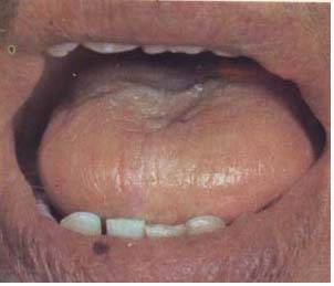
3. Tongue Sores
One or more superficial yellow-white ulcers appear on the tongue, surrounded by congestion, with pain, referred to as “tongue sores”. This condition occurs intermittently and is often caused by excess fire from yin deficiency, heart fire, excessive stomach heat, qi deficiency, or yang deficiency. It is easily triggered after fatigue.
4. Heavy Tongue
The sublingual blood vessels are swollen, resembling another small tongue overlapping the main tongue, referred to as a heavy tongue. This is often caused by heart fire rising or external evil stirring heart fire.
5. Tongue Cancer
The edge of the tongue develops fungoid malignant tissue, with ulceration and pain, producing extremely foul saliva. This is caused by heart and spleen fire stagnation, with qi stagnation, and is classified as a malignant lesion of the tongue.
6. Tongue Fungus (Tongue Papilloma)
A yellow bean-sized growth appears on the tongue, painless, with slow growth and no ulceration, indicating a better prognosis.
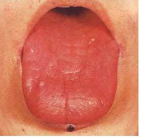
7. Tongue Hemangioma
This image shows a congenital tongue hemangioma. There is a mass on the tongue, slightly purple in color, with no discomfort. This is caused by stagnation of blood vessels in the tongue, with no diagnostic significance, and should be differentiated from cyanotic swollen tongue.
8. Tongue Ulcer (Tongue Cancer)
This condition commonly occurs on the middle and back parts of the tongue, initially presenting as a small ulcer on the tongue edge, gradually enlarging, causing pain that hinders eating, and if it ulcerates and involves blood vessels, it can lead to severe bleeding.
9. Tongue Bleeding
Bleeding from the tongue is referred to as “tongue bleeding”. It can be caused by heart channel heat stagnation, spleen and kidney deficiency fire rising, etc. Generally, bleeding caused by excess fire is more profuse, often accompanied by swelling and pain of the tongue; bleeding caused by deficiency fire is more likely to show oozing blood without swelling of the tongue. It can also be caused by qi not holding blood.