In clinical practice of Traditional Chinese Medicine (TCM), tongue diagnosis is an important method of observation. After examining the surface of the tongue, the doctor may say, “Let’s take a look at the underside of the tongue.” Patients might find this strange, wondering what is being examined. Today, let us explore the sublingual collaterals.
What are the Sublingual Collaterals?
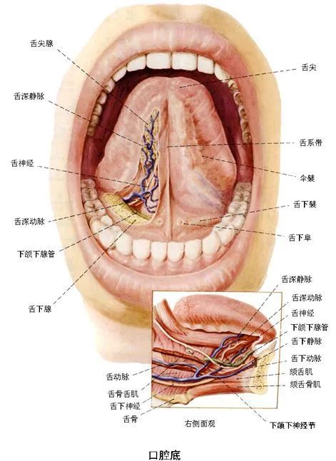
The sublingual collaterals refer to the blood vessels located on both sides of the lingual frenulum, accompanied by the course of the lingual nerve. Currently, it is widely believed that the lingual vein is the primary focus in the diagnosis of the sublingual collaterals.
01
The concept of the sublingual collaterals first appeared in the Huangdi Neijing (Yellow Emperor’s Inner Canon), where it is referred to as 舌下两脉 (sublingual two vessels), 足少阴舌下 (sublingual of the Foot Shaoyin), and 廉泉 (Lianquan). The Suwen (Plain Questions) states, “The sublingual two vessels are Lianquan.”
The Ling Shu (Spiritual Pivot) records, “The root of the Foot Shaoyin is three inches above the ankle, marked at the back of the foot, which is the sublingual two vessels.”
02
During the Tang Dynasty, Sun Simiao placed great emphasis on tongue diagnosis. In his work Qianjin Yaofang (Essential Prescriptions Worth a Thousand Gold), he included dedicated chapters on tongue theory and tongue diseases, stating, “Just by observing the shape of the sublingual collaterals, one can see forms resembling insects, such as crickets or silkworms, with distinct heads and tails, the head being slightly white…”
Why Observe the Sublingual Collaterals?
In the Qing Dynasty, Zhou Xuehai analyzed the examination of the sublingual collaterals in relation to the circulation of Qi and blood and the functions of the organs. In his work Xingse Waizhen Jianmo (External Diagnosis of Form and Color), he recorded, “When the sublingual collaterals appear withered and lifeless, lacking vitality, it indicates that the organ Qi is not reaching there” (here, ‘there’ refers to the sublingual collaterals).
01
At the same time, Zhou Xuehai proposed that a dilated sublingual collateral with a dark purple tongue body indicates the pathological mechanism of blood stasis obstructing the collaterals.
“If the tongue body appears bluish, it indicates that turbid blood fills the fine collaterals, which are the mysterious chambers. The so-called turbid blood filling means that the blood circulating in the mysterious chambers of the tongue is mixed with turbid Qi. This may be due to cold Qi condensing or phlegm obstructing the meridians of the stomach and pericardium, causing the blood to surge upwards. This does not necessarily mean that the blood has decayed before this occurs.”
02
Modern research indicates that abnormalities such as dilated and tortuous sublingual collaterals, or the appearance of petechiae, are associated with microcirculation disorders, increased venous pressure, abnormal blood rheology, increased blood coagulation, or decreased fibrinolysis, and increased platelet aggregation or hyperfunction of release.
Typical Sublingual Manifestations
Identifying
Sublingual
Manifestations
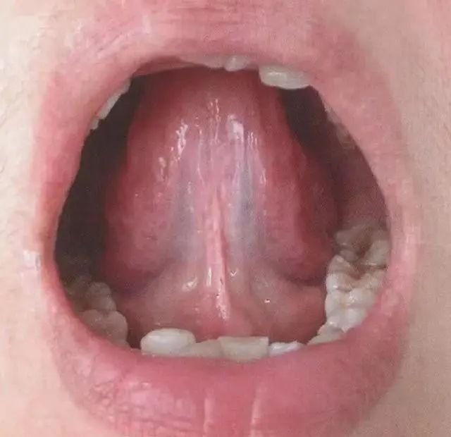
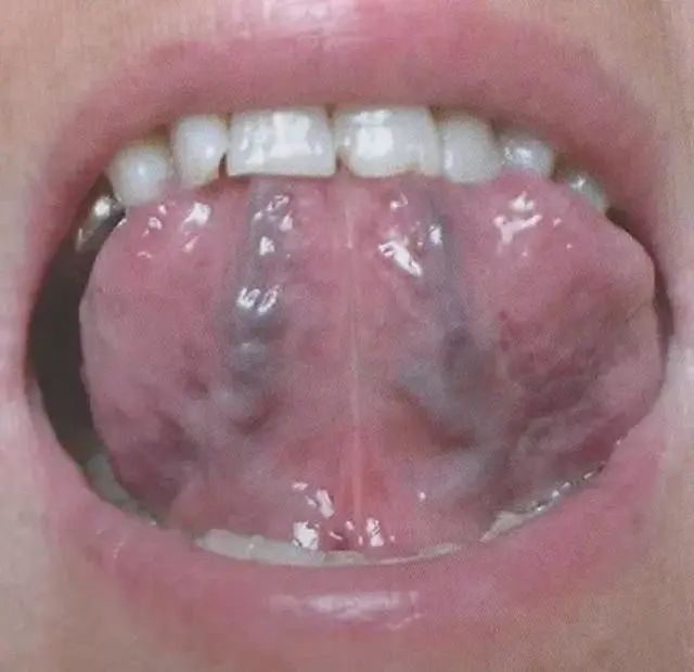
Image|Normal Sublingual Collaterals
The normal sublingual collaterals are located on both sides of the lingual frenulum, running longitudinally, with a diameter of less than 2.7mm, and a length not exceeding 3/5 of the distance from the sublingual tuberosity to the tip of the tongue. The color of the collaterals is light purple.
Image|Thickened Sublingual Vein with Branches
The sublingual vein appears thickened, or purplish spots may be seen, indicating possible blood stasis.
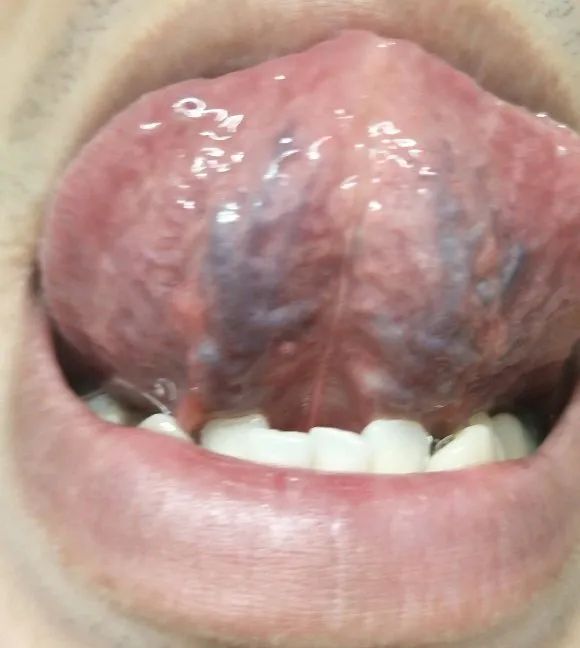
Image|Dilated and Thickened Sublingual Collaterals
The dilated and thickened sublingual collaterals, with a bluish-purple or dark purple color, indicate possible blood stasis.
Warm Reminder
Friends, you can also observe your own tongue in the mirror. If you notice any abnormal tongue manifestations or are uncertain about your tongue appearance, do not panic. You can come to the hospital for consultation to resolve your doubts.
Wang Jianming
MD, Chief Physician, Graduate Supervisor
Deputy Director of the Department of Traditional Chinese Medicine and Rheumatology at the China-Japan Friendship Hospital, Teaching Director
Member of the Rheumatology Branch of the China Association of Chinese Medicine
Member of the Rheumatology Branch of the Chinese Association of Integrative Medicine
Executive Director of the Rheumatology Branch of the World Federation of Chinese Medicine Societies
Executive Director of the Rheumatology Branch of the Chinese Ethnic Medicine Society
Review Expert for the National Natural Science Foundation of China
Academic Guidance Expert for the Rheumatology Community of Beijing-Tianjin-Hebei
Expert in Medical Accident Appraisal for the Beijing Medical Association
Guidance Teacher for the Grassroots TCM Experts and Academic Experience Inheritance Project in Chaoyang District, Beijing
Specializes in using TCM and integrative methods to treat various rheumatic diseases, such as rheumatoid arthritis, ankylosing spondylitis, fibromyalgia syndrome, gout, Sjögren’s syndrome, postpartum rheumatism, osteoarthritis, psoriatic arthritis, Behçet’s disease, systemic lupus erythematosus, myositis/dermatomyositis, vasculitis, scleroderma, etc.
Outpatient Hours
●China-Japan Friendship Hospital 2nd Floor, Building M, Department of Traditional Chinese Medicine and Rheumatology
Monday morning, Room 24
Thursday afternoon, Room 26
●Beijing TCM Hospital Shunyi Hospital Rheumatology Department (Currently, Shunyi outpatient service is suspended due to the pandemic, resumption time to be determined)
Monday afternoon (every other week)

More Health Education
Welcome to follow us by scanning the QR code
WeChat Official Account | Wang Jianming Rheumatology Forum