Sublingual collaterals are longitudinal veins located on both sides of the lingual frenulum, primarily reflecting the state of qi and blood circulation. Common abnormal manifestations of the sublingual collaterals include: coarse and long veins resembling a net, varicose veins, and blood stasis in the veins.
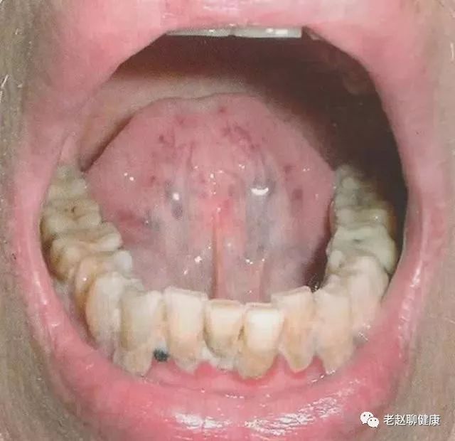
Normal sublingual veins:
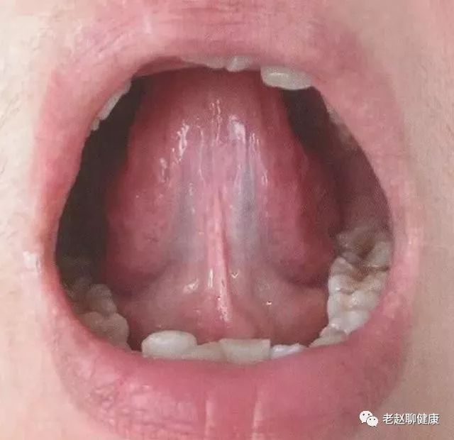
Characteristics of the tongue:
The sublingual collaterals are large collaterals located longitudinally on both sides of the lingual frenulum, with a diameter of less than 2.7mm. Their length does not exceed 3/5 of the distance from the sublingual tubercle to the tip of the tongue, and the color of the collaterals is light purple.
Clinical significance:
Normal sublingual veins.
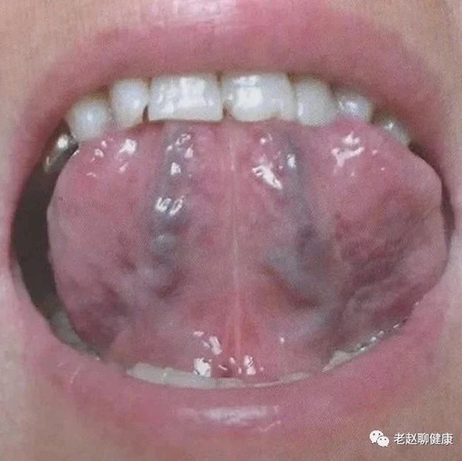
Coarse and long veins resembling a net:
Characteristics of the tongue:
The sublingual collaterals are swollen and elongated, appearing purple or purplish-black in a net-like pattern.
Clinical significance:
This is a sign of qi and blood stasis.
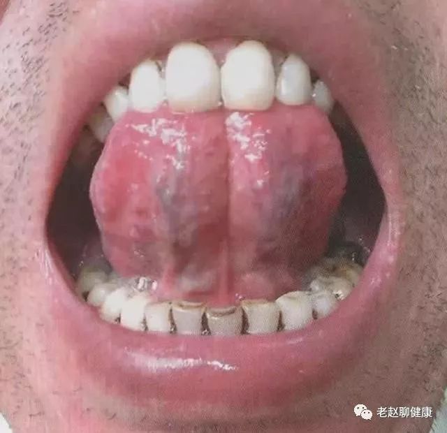
Varicose veins:
Characteristics of the tongue:
The sublingual collaterals are significantly varicose and thickened, with a color of bluish-purple or dark purple.
Clinical significance:
This is often caused by qi stagnation and blood stasis, leading to obstructed blood flow.
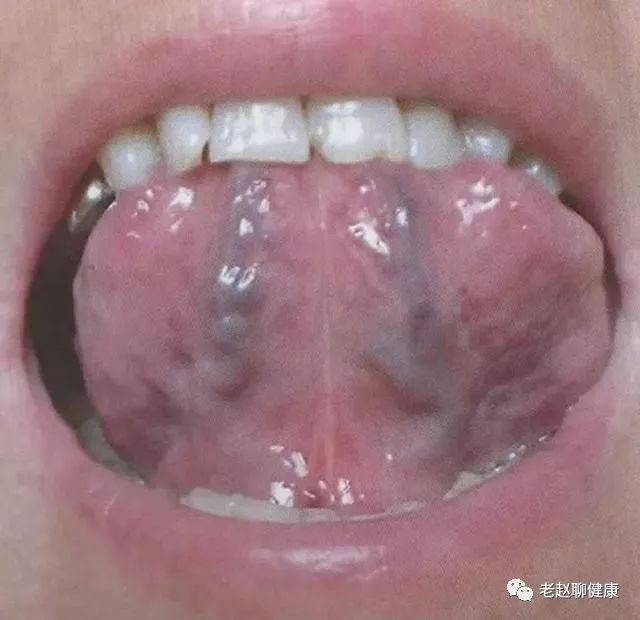
Blood stasis in the veins:
Characteristics of the tongue:
The sublingual collaterals or small veins appear bluish-purple or purplish-black, or the collaterals show changes such as uneven purple blood stasis nodules.
Clinical significance:
This is a sign of blood stasis, which can be caused by various factors such as cold blood, hot blood, qi stagnation, phlegm-dampness, and yang deficiency.